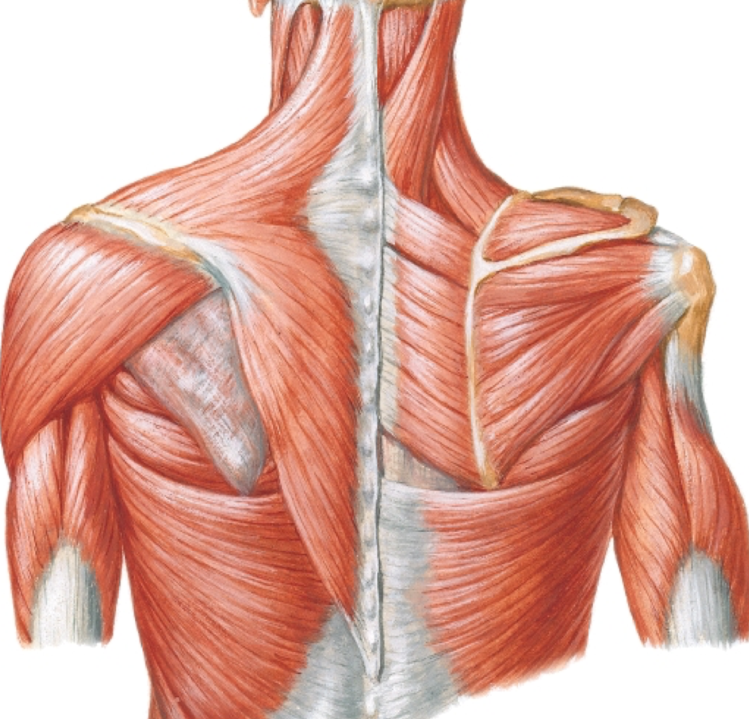
Shoulder Muscles Diagram Back / Back Shoulder Muscle Anatomy Human Anatomy / Tutorials on the shoulder muscles (e.g rotator cuff muscles:. The shoulder muscles produce the characteristic shape of the shoulder and can be classified into two groups: The core muscles are those in the abdomen, back, and pelvis, and they also stabilize the body and assist in tasks, such as lifting weights. The muscles of the superficial layer of the back move the shoulder blade (scapula) and upper arm (humerus). The deltoid, teres major, teres minor, infraspinatus, supraspinatus (not shown) and subscapularis muscles (not shown) all extend from the scapula to the humerus and act on the shoulder joint. Lateral) • muscles of the hand.
The latissimus dorsi muscle spans from the lower back to the upper arm and is partially covered by the trapezius. The anterior deltoid, the lateral deltoid, and the posterior deltoid. Мышцы гипотенара мышцы тенара hypothenar muscules thenar muscules. Tutorials on the shoulder muscles (e.g rotator cuff muscles: Stability is mostly offered by the periarticular muscles, that originate from the scapula and insert on the caput humeri.
Shoulder Anatomy Back Anatomy Drawing Diagram from d1yboe6750e2cu.cloudfront.net The shoulder muscles bridge the transitions from the torso into the head/neck area and into the upper extremities of the arms and hands. I started this website back in late 2009 during college, and it has been my pet project ever since. Shoulder anatomy images shoulder muscle tissues anatomy actions diagram. It is opposite from the chest, and the vertebral column runs the best way to strengthen back muscles is in a static position. How to build a wide back. The following diagram shows all the major back muscles. Shoulder muscles back muscles shoulder muscle anatomy neck muscle anatomy shoulder joint chest muscles major muscles bones and muscles what are the most beneficial back exercises? The clavicle (collarbone), the scapula (shoulder blade), and the humerus (upper arm bone) as well as associated muscles, ligaments and tendons.
This flow diagram provides an aid to diagnosis of shoulder conditions
Deadlift muscles will include knee, hip, and back extensors, which primarily include the quads, glutes, and spinal erectors. Now i am training to correct both my muscular imbalance and right shoulder issues. As one of the four muscles of the rotator cuff, the main function is to externally rotate the humerus and stabilize the shoulder joint. This rotator cuff includes the m.supraspinatus, m as the disease progresses, night pain becomes more common. Names and diagram | science trends there are three different muscle groups found in the back: The shoulder muscles bridge the transitions from the torso into the head/neck area and into the upper extremities of the arms and hands. The superficial group, the deep group, and the intermediate group. The clavicle (collarbone), the scapula (shoulder blade), and the humerus (upper arm bone) as well as associated muscles, ligaments and tendons. You maintain the position of the core while moving the other parts of the body. Start studying back & shoulder muscles. Human anatomy diagrams show internal organs, cells, systems, conditions, symptoms and sickness information and/or tips for healthy living. Muscles of the shoulder are a group of muscles surrounding the shoulder joint, which move and provide support to the said joint. Muscle tendons stretch over joints and contribute to joint stability.
The trapezius and latissimus dorsi muscles connect the upper limb to the vertebral column. Shoulder anatomy images shoulder muscle tissues anatomy actions diagram. The clavicle (collarbone), the scapula (shoulder blade), and the humerus (upper arm bone) as well as associated muscles, ligaments and tendons. The human back extends from the buttocks to the posterior portion of the neck and shoulders. Other muscles are small and cover much less space.
Do You Want A Wide Thick And Strong Back Then Here We Have Just The Exercises And Workouts For You Yoga Anatomy Muscle Anatomy Human Anatomy And Physiology from i.pinimg.com This diagram depicts back shoulder muscles. Shoulder anatomy images shoulder muscle tissues anatomy actions diagram. The core muscles are those in the abdomen, back, and pelvis, and they also stabilize the body and assist in tasks, such as lifting weights. Lateral) • muscles of the hand. Muscles of the forearm (posterior view). Certain back muscles extend to other areas, like the shoulders, upper arms, and thighs. It is opposite from the chest, and the vertebral column runs the best way to strengthen back muscles is in a static position. The anterior deltoid, the lateral deltoid, and the posterior deltoid.
There are three main muscles in your shoulder:
Deadlift muscles will include knee, hip, and back extensors, which primarily include the quads, glutes, and spinal erectors. It is opposite from the chest, and the vertebral column runs the best way to strengthen back muscles is in a static position. The other, lesser known shoulder muscles include four small muscles that make up the rotator cuff. Weak muscle tissues are amongst the prime causes of back discomfort, especially in the decrease back. Wrist extension, by contrast, shortens the angle at the back of the wrist. The next life study seated female figure, shows the upper part of the pectoralis major positioned flat against the rib cage, with very little thickness. The superficial group, the deep group, and the intermediate group. The muscles of the superficial layer of the back move the shoulder blade (scapula) and upper arm (humerus). Human anatomy diagrams show internal organs, cells, systems, conditions, symptoms and sickness information and/or tips for healthy living. Although three ligaments protect and surround the shoulder joint, most of its stability comes from the powerful muscles and tendons of the rotator cuff. As one of the four muscles of the rotator cuff, the main function is to externally rotate the humerus and stabilize the shoulder joint. Facebook twitter whatsapp pin it. Start studying back & shoulder muscles.
Shoulder muscles back muscles shoulder muscle anatomy neck muscle anatomy shoulder joint chest muscles major muscles bones and muscles what are the most beneficial back exercises? There are three main muscles in your shoulder: Human anatomy diagrams show internal organs, cells, systems, conditions, symptoms and sickness information and/or tips for healthy living. Shoulder anatomy images shoulder muscle tissues anatomy actions diagram. Мышцы гипотенара мышцы тенара hypothenar muscules thenar muscules.
Deltoid Muscle Wikipedia from upload.wikimedia.org Tutorials on the shoulder muscles (e.g rotator cuff muscles: Start studying back & shoulder muscles. Names and diagram | science trends there are three different muscle groups found in the back: This diagram depicts back shoulder muscles. Muscle tendons stretch over joints and contribute to joint stability. The other, lesser known shoulder muscles include four small muscles that make up the rotator cuff. This flow diagram provides an aid to diagnosis of shoulder conditions The superficial group, the deep group, and the intermediate group.
Muscle tendons stretch over joints and contribute to joint stability.
Shoulder stretches are necessary to maintain a balance among the muscles around the shoulders and upper back. Although three ligaments protect and surround the shoulder joint, most of its stability comes from the powerful muscles and tendons of the rotator cuff. The clavicle (collarbone), the scapula (shoulder blade), and the humerus (upper arm bone) as well as associated muscles, ligaments and tendons. The anterior deltoid, the lateral deltoid, and the posterior deltoid. Human anatomy diagrams show internal organs, cells, systems, conditions, symptoms and sickness information and/or tips for healthy living. This diagram depicts back shoulder muscles. It is opposite from the chest, and the vertebral column runs the best way to strengthen back muscles is in a static position. Shoulder muscles back muscles shoulder muscle anatomy neck muscle anatomy shoulder joint chest muscles major muscles bones and muscles what are the most beneficial back exercises? Lateral) • muscles of the hand. Basic muscle anatomy 12 photos of the basic muscle anatomy basic muscle anatomy, basic muscle anatomy and physiology crossword puzzle answers, basic muscle anatomy diagram, basic muscle anatomy pdf, major muscle groups anatomy, human muscles, basic muscle anatomy. Tutorials on the shoulder muscles (e.g rotator cuff muscles: The shoulder muscles produce the characteristic shape of the shoulder and can be classified into two groups: The deltoid, teres major, teres minor, infraspinatus, supraspinatus (not shown) and subscapularis muscles (not shown) all extend from the scapula to the humerus and act on the shoulder joint.
The clavicle (collarbone), the scapula (shoulder blade), and the humerus (upper arm bone) as well as associated muscles, ligaments and tendons shoulder muscles diagram. The deltoid, teres major, teres minor, infraspinatus, supraspinatus (not shown) and subscapularis muscles (not shown) all extend from the scapula to the humerus and act on the shoulder joint.


0 Komentar